If you have been recommended for any type of radiation therapy to treat cancer, your care team will request a scan, such as a CT or an MRI, before you start your course of treatment. The purpose of this staging scan is to determine the exact location, size and behaviour of the tumour to help plan your radiotherapy.
During proton beam therapy, radiation is directed at cancer cells, where it stops, avoiding healthy cells. This level of precision makes medical imaging, like CT and MRI, essential to PBT. It creates a 3-D image of the affected site, enabling radiation oncologists to create an accurate, fully bespoke treatment plan.
Read on to discover the common types of medical imaging and how they play a vital role in treating cancer with radiation therapy, particularly proton beam therapy.
Understanding how proton beam therapy works
Proton beam therapy delivers a carefully controlled beam of energy that stops right at the tumour, rather than passing through the body. This means doctors can treat cancer with great precision, helping to protect healthy tissue and reduce common side effects. PBT is available at our private treatment centre at UCLH, London.
Read more about how proton beam therapy works.
Why imaging matters in proton beam therapy cancer treatment
Imaging is essential at every stage of radiation treatment, including proton beam therapy. Scans enable your team to plan, deliver and monitor your treatment with great accuracy and safety.
Here is how medical imaging is used throughout the proton treatment process:
Before treatment
Detailed scans, such as CT or MRI, are used to map the exact size, shape, and position of the tumour, and to assess how it interacts with normal tissue and organs. A few weeks before your treatment is due to start, you will be invited to have CT and/or MRI scans.
These planning scans are used by the radiotherapy team to create a personalised treatment plan. They will accurately outline the tumour and a margin around it, which also needs to be treated. Then radiation can be precisely aimed where it’s needed, sparing normal cells.
During treatment
Once in the treatment room, radiographers will place you carefully in the right position on a bed beneath the proton beam therapy machine. Image-guided radiation therapy (IGRT), a process in which CT scans or X-rays are captured before treatment, is used to confirm your position and allow for fine adjustments as needed. Even small movements or anatomical shifts can affect the path of the proton beam, so this part of the process is essential.
You may be required to wear an immobilisation device to help you stay perfectly still during radiotherapy. For example, head masks for the head and neck area and body moulds for the spine and trunk. This ensures that the treatment is delivered safely and accurately, maintaining the exact same position for your daily radiation treatments.
After treatment
Follow-up scans in the months after treatment help the clinical team monitor how the tumour is responding to treatment and to check for any treatment-related changes in the surrounding tissues.
Types of imaging used in proton beam therapy
CT scans (Computerised Tomography)
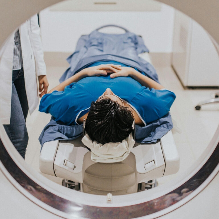
CT is a common type of scan used during proton beam therapy treatment. X-rays are used to create detailed 3D images of the treatment area, helping the radiotherapy team see the exact size and shape of the tumour. It is also used for ongoing verification and to see how the tumour is reacting to treatment.
CT scans are quick and painless, taking only a few minutes. You’ll be asked to lie still on a table that moves through a doughnut-shaped scanner, which is not enclosed. While the scan involves a small amount of radiation, the dose is carefully controlled and kept as low as possible to minimise any risk.
MRI scans (Magnetic Resonance Imaging)
MRI scans provide excellent soft-tissue contrast, giving doctors a clearer view of complex structures that may not be as clearly visible on CT. Although MRI is less commonly used for real-time treatment verification, it can be invaluable during planning, particularly for tumours near critical organs or soft tissue.
An MRI scan takes longer than a CT. You will pass through a tunnel-like machine, and the scan sequences can be loud and noisy. While it might feel confined, it’s completely non-invasive and uses magnets rather than radiation.
PET scans (Positron Emission Tomography)
PET scans identify a tumour’s metabolic activity, indicating how active the cancer is, whether it has spread to local lymph nodes or other organs. A PET scan is often combined with a CT scan to offer both structural and functional detail.
During a PET scan, you are injected with a small safe amount of radioactive tracer, which is taken up in the tumour, meaning that it is seen more clearly on the scan. The scanner is similar to a CT scanner and is not enclosed.
Find out more about proton beam therapy today
At Proton International London, we deliver private proton beam therapy to patients from around the world at our centre in the highly respected University College London Hospitals (UCLH). Our clinicians work with highly skilled imaging specialists and use state-of-the-art technologies to ensure every patient benefits from the safest and most accurate proton beam therapy possible.
To find out if proton beam therapy is a suitable treatment for you or your loved one, explore our site for more information about the cancers we treat and what the benefits are. Talk to your consultant oncologist in the first instance, and if they think you are suitable, they can refer you for private therapy. In the meantime, if you have a question, please do not hesitate to contact us.
Conclusion
Imaging is essential to every stage of proton beam therapy, from staging a tumour to planning and treatment delivery, and post-treatment monitoring. Techniques such as CT, MRI, and PET scans allow clinicians to visualise the tumour in three dimensions and ensure the proton dose is delivered with high accuracy.
Frequently asked questions about the use of imaging for proton beam therapy
Why is MRI preferred for some cancers over CT in planning proton beam therapy?
MRI is often recommended alongside CT for cancers that occur in soft tissues, such as sarcomas and prostate cancer. It provides a high contrast image of the tissue, helping to define the tumour’s boundaries. A CT is still essential for calculating the radiation dosage required.
Does imaging add extra radiation exposure?
CT and PET scans use ionising radiation. While this does contribute to overall radiation exposure, the dosage is carefully managed, and the benefit almost always outweighs the potential risk. CT and other nuclear medicine tests involve higher doses than X-rays, so the need for repeated imaging is carefully assessed, particularly in children and young adults.
Can imaging happen during proton beam therapy sessions?
During proton beam therapy, image-guided radiotherapy (IGRT) is carried out to verify the tumour’s position before and during treatment. This allows for fine adjustments to maximise precision and to protect surrounding healthy tissue.
How does imaging improve dose calculation in proton beam therapy?
Imaging significantly enhances the accuracy of proton dose calculation by providing detailed anatomical information that guides the beam’s delivery. In traditional radiotherapy, high-energy X-rays are used, which pass continuously through the body, exposing healthy cells to radiation.
In proton beam therapy, most of the radiation is deposited at the tumour site and then stops almost completely. That’s why accurate imaging is so important to ensure that the radiation dose is delivered to the precise tumour location.